Plateforme de Microscopie
Responsable : Emmanuelle BLANCHARD-LAUMONNIER
Plateforme IBiSA de Microscopie Electronique de l’Université de Tours.
Le Département des Microscopies est un plateau technique mutualisant les appareils et les compétences d’imagerie microscopique au service des équipes de l’Université de Tours, des grands organismes extérieurs à l’Université et du secteur privé industriel.
Ce département est associé à plusieurs structures fédératives : PST Analyse des Systèmes Biologiques, Groupement de Moyens Scientifiques et Techniques de Tours (GMSTT), FED 4225 (ancien IFR 136) et FED 4226 (ancien IFR 135). Il est membre du Réseau d’Imagerie en Microscopie Electronique (RIME, anciennement RCCM).
La plate-forme a été labellisée IBiSA (Infrastructures en Biologie Santé et Agronomie) en 2015.
Ce département est associé à la l’US-61 Analyse des Systèmes Biologiques
Prestations
Prestations internes
Elles constituent un support logistique indispensable aux deux FED de l’Université (FED 4225 « Agents Infectieux, Immunité et Thérapies » et FED 4226 « Neuroimagerie Fonctionnelle: de l’image à la fonction ») ainsi qu’aux autres équipes de l’axe Sciences de la Vie et de la Santé et de l’axe Sciences et Technologies.
Les prestations peuvent aller de la seule mise à disposition de façon autonome des appareils lourds, jusqu’à la prise en charge complète d’une problématique de recherche spécifique par le personnel encadrant du plateau technique.
Le coût des travaux effectués pour les équipes de Tours est à leur charge selon une tarification propre disponible à leur demande.
Prestations externes
La reconnaissance à l’échelon national et international de la qualité des compétences de ce plateau technique, contribue à développer une politique d’ouverture et de valorisation de l’Université de Tours vers un partenariat public ou privé.
Des collaborations sont établies avec de grands organismes publics extérieurs à l’Université (Institut Pasteur, Museum d’Histoire Naturelle de Paris, équipes INSERM, Necker, St Louis… )
Des prestations de haut niveau sont développées avec le secteur privé industriel national et international, dans tous les domaines d’exploration microscopique.
Ces prestations sont facturées selon un devis établi après étude des projets scientifiques et/ou protocoles techniques et concertation entre les partenaires.
Charte de la Plate-Forme
Définition
Une plate-forme est le regroupement sur un même site des équipements et des moyens humains destinés à offrir à une communauté d’utilisateurs des ressources technologiques de haut niveau. En adoptant la présente charte, la plate-forme s’engage à une ouverture large au niveau régional et national, non seulement aux équipes du site, mais aussi aux expérimentateurs extérieurs, quel que soit leur rattachement (organismes publics, entreprises…). La reconnaissance d’une plate-forme et les moyens spécifiques (financement, personnel) qui en découlent sont conditionnés par un cahier des charges général, qui pourra être précisé selon la thématique de la plate-forme.
Ouverture
La plate-forme devant être ouverte aux équipes extérieures au site, les locaux qui lui sont affectés doivent présenter une superficie suffisante pour accueillir les équipes utilisatrices. Cette ouverture impose des effectifs dédiés à la plate-forme qui ont la capacité de répondre à la demande de service et d’encadrement.
Mode de gestion
Le fonctionnement et l’animation de la plate-forme sont sous la responsabilité d’une personne bien identifiée. La plate-forme se dote d’un conseil scientifique, comportant des utilisateurs et des experts extérieurs, qui définit les priorités des projets, s’assure de la qualité des prestations offertes et propose les évolutions méthodologiques. La plate-forme affiche de manière formalisée l’offre de service, les conditions d’accès et les tarifs.
Pour le fonctionnement de la plate-forme, le GIS IBiSA conseille la mise en place d’un management de la qualité inspiré par la norme ISO 9001. Si une démarche de certification ISO 9001 peut être une aide à l’optimisation des activités des PF voire à leur valorisation, elle ne peut en aucun cas constituer un critère d’évaluation scientifique et technologique pour obtenir la labellisation IBiSA.
Evolution technologique
La plate-forme doit assurer la pérennité de sa performance technologique par une veille technologique active et un plan d’action pour la mise à niveau des matériels et la formation des personnels. Cet objectif est d’autant mieux atteint que la plate-forme soutient des recherches à caractère méthodologique aboutissant à des publications scientifiques ou à des actions de valorisation (dépôt de brevet, création de jeunes pousses…).
Formation
Les ressources humaines et matérielles disponibles sur la plate-forme sont mises à profit pour la formation d’étudiants (stagiaires, doctorants…), d’ingénieurs et techniciens et pour des programmes d’animation et de formation scientifiques et techniques (séminaires, ateliers, sessions d’études…).
Evaluation
Le respect des directives énoncées dans le cahier des charges doit être évalué pour chaque plate-forme par une instance mandatée par les organismes ayant procédé à l’affectation des moyens spécifiques. Le degré d’ouverture de la plate-forme aux équipes extérieures est un critère majeur d’évaluation. Cette évaluation a posteriori sera effectuée périodiquement et conditionnera la poursuite du financement ou le maintien du personnel dédié.
(Charte des Plateformes IBiSA)
Compétences
La plate-forme met à disposition les appareils lourds et les compétences scientifiques et techniques des responsables et du personnel encadrant dans tous les domaines de la Microscopie.
Analyse en MET de cellules et tissus et immunomicroscopie électronique
Analyse en MET et MEB de particules (bactéries, virus, ADN, liposomes, macromolécules)
État de surface, analyse élémentaire et cartographie spectrale par MEB et sonde EDS
Reconstruction 3D à partir de coupes sériées en MET
Localisation d’éléments cellulaires marqués (protéines virales, facteurs de signalisation…) par microscopie confocale à balayage laser
Reconstruction 3D des volumes et quantification de la colocalisation de marqueurs à partir d’acquisitions en microscopie confocale
Acquisitions confocales en time-lapse sur cellules vivantes
Equipement
- Microscope Electronique à Transmission JEOL JEM-1400 Plus (120 kV)
- Microscope Electronique à Transmission JEOL JEM-1011 (100 kV)
- Microscope Electronique à Balayage à Effet de Champ Zeiss ULTRA Plus
Equipements de préparation des échantillons
- Ultramicrotomes LEICA Ultracut UCT et UC7
- Cryo-Ultramicrotome LEICA EM UC7/FC7
- Système de congélation haute pression (HPF) LEICA ICE
- Appareil de cryo-substitution LEICA EM AFS2
- Automate de vitrification (cryo-plunge) THERMO Vitrobot
- Appareil à pulvérisation cathodique JEOL JUC-5000
- Métalliseur GATAN Precision Etching Coating System (PECS)
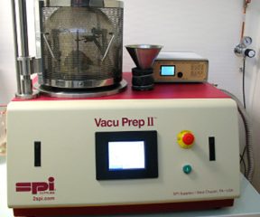
- Evaporateur SPI VacuPrep II
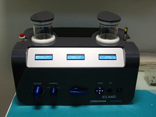
- Station d’effluvage (glow discharge) CORDOUAN Elmo
Microscopie Photonique
Personnels
Le département est animé par une équipe encadrante d’une dizaine de personnes :
Un responsable administratif et scientifique : Philippe ROINGEARD (PU-PH)
Une secrétaire-gestionnaire : Isabelle SAUSSEREAU
2 ingénieurs de recherche : Pierre-Ivan RAYNAL et Rustem UZBEKOV
1 ingénieur d’études : Julien BURLAUD-GAILLARD
2 techniciennes : Juliette ROUSSEAU et Emma PERDRIAU
Il est localisé dans les locaux du pôle de Biologie du CHU Bretonneau auquel appartient l’Unité Fonctionnelle de Biologie Cellulaire du service d’Anatomie Pathologique.
Cette unité composée de 6 personnes est dirigée par le Pr Philippe ROINGEARD et développe deux types d’activités, dont l’une est utilisatrice du département des Microscopies :
- une activité de Pathologie Ultra-Structurale : Emmanuelle BLANCHARD-LAUMONNIER (PU-PH) et Sébastien EYMIEUX (PHU)
- une activité de Biologie Moléculaire : Philippe ROINGEARD (PU-PH) et Christophe HOURIOUX (PU-PH)
Les responsables sont assistés par deux techniciennes : Marie-Paule REGNER et Florentine CHESNEL.
Composition de l’équipe :
- Responsable administratif et scientifique, PU-PH : Philippe ROINGEARD
- Secrétaire-gestionnaire : Isabelle SAUSSEREAU
- Responsable technique, Ingénieur de Recherche : Pierre-Ivan RAYNAL
- Ingénieur de Recherche : Rustem UZBEKOV
- Ingénieur d’Etudes : Julien BURLAUD-GAILLARD
- Techniciennes : Juliette ROUSSEAU et Emma PERDRIAU
- Unité Fonctionnelle de Biologie Cellulaire :
- – Pathologie Ultra-structurale
- PU-PH : Emmanuelle BLANCHARD-LAUMONNIER
- PHU : Sébastien EYMIEUX
- Technicienne : Florentine CHESNEL – Biologie Moléculaire
- PU-PH : Christophe HOURIOUX
- Technicienne : Marie-Paule REGNER
Le laboratoire a été fondé par le Dr Brigitte ARBEILLE (1947-2015).
Travaux & Collaborations
Répartitions des secteurs d’activité
- Secteur académique local : 39 %
- Secteur académique extérieur : 27 %
- Secteur privé : 34 %
(période 2014-2018)
Publications
Travaux publiés dans des revues internationales à comité de lecture ayant fait l’objet de prestations du Département des Microscopies de la PST ASB / Plateforme IBiSA de Microscopie Electronique de l’Université de Tours
(période 2016–2020)
Les noms des membres du Département des Microscopies sont soulignés.
- Equipes de l’université de Tours – Secteur Biologie & Santé
- Equipes de l’université de Tours – Secteur Sciences des Matériaux
- Equipes nationales
- Equipes internationales
- Publications de la plateforme en partenariat avec des équipes de recherche ou des industriels
Equipes de l’université de Tours – Secteur Biologie & Santé
UMR INSERM U1069 : Nutrition, Croissance et Cancer (N2C)
- GAMBADE A, ZREIKA S, GUEGUINOU M, CHOURPA I, FROMONT G, BOUCHET AM, BURLAUD-GAILLARD J, POTIER-CARTEREAU M, ROGER S, AUCAGNE V, CHEVALIER S, VANDIER C, GOUPILLE C, WEBER G.
Activation of TRPV2 and BKCa channels by the LL-37 enantiomers stimulates calcium entry and migration of cancer cells.
Oncotarget. 2016 ; 7(17) : 23785-23800. - BON E, DRIFFORT V, GRADEK F, MARTINEZ-CACERES C, ANCHELIN M, PELEGRINP, CAYUELA ML, MARIONNEAU-LAMBOT S, OULLIER T, GUIBON R, FROMONT G, GUTIERREZ-PAJARES JL, DOMINGO I, PIVER E, MOREAU A, BURLAUD-GAILLARDJ, FRANK PG, CHEVALIER S, BESSON P, ROGER S.
SCN4B acts as a metastasis-suppressor gene preventing hyperactivation of cell migration in breast cancer.
Nat Commun. 2016 ; 7 : 13648.
UMR INSERM U1100 : Centre d’Etude des Pathologies Respiratoires (CEPR)
- HAMON Y, LEGOWSKA M, HERVE V, DALLET-CHOISY S, MARCHAND-ADAM S, VANDERLYNDEN L, DEMONTE M, WILLIAMS R, SCOTT C, SI-TAHAR M.
Neutrophilic cathepsin C is matured by a multistep proteolytic process and secreted by activated cells during inflammatory lung diseases.
J Biol Chem. 2016 ; 291(16) : 8486-8499. - CHEVALEYRE C, RIOU M, BREA D, VANDEBROUCK C, BARC C, PEZANT J, MELO S, OLIVIER M, DELAUNAY R, BOULESTEIX O, BERTHON P, ROSSIGNOL C, BURLAUD-GAILLARD J, BECQ F, GAUTHIER F, SI-TAHAR M, MEURENS F, BERRI M, CABALLERO-POSADAS I, ATTUCCI S.
The Pig: A Relevant Model for Evaluating the Neutrophil Serine Protease Activities during Acute Pseudomonas aeruginosa Lung Infection.
PLoS One. 2016 ; 11(12) : e0168577. - KRYZA T, PARENT C, PARDESSUS J, PETIT A, BURLAUD-GAILLARD J,REVERDIAU P, IOCHMANN S, LABAS V, COURTY Y, HEUZE-VOURC’H N.
Human kallikrein-related peptidase 12 stimulates endothelial cell migration by remodeling the fibronectin matrix.
Sci Rep. 2018 ; 8(1): 6331. - GUILLON A, FOUQUENET D, MORELLO E, HENRY C, GEORGEAULT S,
SI-TAHAR M, HERVÉ V.
Treatment of Pseudomonas aeruginosa biofilm present in endotracheal tubes by poly-L-Lysine.
Antimicrob Agents Chemother. 2018 ; 62(11). - WARTENBERG M, SAIDI A, GALIBERT M, JOULIN-GIET A, BURLAUD-GAILLARDJ, LECAILLE F, SCOTT CJ, AUCAGNE V, DELMAS AF, LALMANACH G.
Imaging of extracellular cathepsin S activity by a selective near infrared fluorescence substrate-based probe.
Biochimie. 2019 ; 166 : 84-93 - BERNARD L, DESOUBEAUX G, BODIER-MONTAGUTELLI E, PARDESSUS J, BREA D, ALLIMONNIER L, EYMIEUX S, RAYNAL P.I, VASSEUR V, VECELLIOL, MATHE L, GUILLON A, LANOTTE P, POURCHEZ J, O VERHOEVEN P, ESNOUF S, FERRY M, ETERRADOSSI N, BLANCHARD Y BROWN P, ROINGEARD P, ALCARAZJ.P, CINQUIN P, SI-TAHAR M, HEUZE-VOURC’H N.
Controlled heat and humidity-based treatment for the re-use of protective face-masks : a pragmatic proof-of-concept to address the mass shortage of face-masks and prevent the SARS-CoV2 transmission.
Front Med. 2020 ; 7 : 584036.
UMR INSERM U1253 : Imagerie et Cerveau (iBrain)
- ZEMMOURA I, BLANCHARD E, RAYNAL PI, ROUSSELOT-DENIS C, DESTRIEUX C, VELUT S.
How Klingler’s dissection permits exploration of brain structural connectivity? An electron microscopy study of human white matter.
Brain Struct Funct. 2016 ; 221(5) : 2477-2486. - DANGOUMAU A, MAROUILLAT S, GAILLARD J, UZBEKOV R,VEYRAT-DUREBEX C, BLASCO H, ARNOULT C, CORCIA P, ANDRES C, VOURC’H P.
Inhibition of pathogenic mutant SOD1aggregation in cultured motor neuronal cells by prevention of its SUMOylation on lysine 75.
Neurodegener Dis. 2016 ; 16(3-4) : 161-171. - UNG DC, IACONO G, MEZIANE H, BLANCHARD E, PAPON MA, SELTEN M, VAN RHIJN JR, MONTJEAN R, RUCCI J, MARTIN S, FLEET A, BIRLING MC, MAROUILLAT S, ROEPMAN R, SELLOUM M, LUX A, THEPAULT RA, HAMEL P, MITTALK, VINCENT JB, DORSEUIL O, STUNNENBERG HG, BILLUART P, NADIF KASRI N, HERAULT Y, LAUMONNIER F.
Ptchd1 deficiency induces excitatory synaptic and cognitive dysfunctions in mouse.
Mol Psychiatry. 2018 ; 23 : 1356-1367.
UMR INSERM U1259 : Morphogénèse et Antigénicité du VIH et du Virus des Hépatites (MAVIVH)
- MONEL B, COMPTON AA, BRUEL T, AMRAOUI S, BURLAUD-GAILLARD J,ROY N, GUIVEL-BENHASSINE F, PORROT F, GENIN P, MEERTENS L, SINIGAGLIA L, JOUVENET N, WEIL R, CASARTELLI N, DEMANGEL C, SIMON-LORIERE E, MORIS A,
ROINGEARD P, AMARA A, SCHWARTZ O.
Zika virus induces massive cytoplasmic vacuolization and paraptosis-like death in infected cells.
EMBO J. 2017 ; 36(12):1653-1668. - PIVER E, BOYER A, GAILLARD J, BULL A, BEAUMONT E, ROINGEARD P, MEUNIER JC.
Ultrastructural organisation of HCV from the bloodstream of infected patients revealed by electron microscopy after specific immunocapture.
Gut. 2017, 66: 759-761. - MEYER L, LEYMARIE O, CHEVALIER C, ESNAULT E, MOROLDO M, DA COSTA B,
GEORGEAULT S, ROINGEARD P, DELMAS B, QUERE P, LE GOFFICR.
Transcriptomic profiling of a chicken lung epithelial cell line (CLEC213) reveals a mitochondrial respiratory chain activity boost during influenza virus infection.
PLoS One. 2017 ; 12(4):e0176355. - TARTOUR K, NGUYEN XN, APPOURCHAUX R, ASSIL S, BARATEAU V, BLOYET LM,
BURLAUD-GAILLARD J, CONFORT MP, ESCUDERO-PEREZ B, GRUFFAT H, HONGSS, MOROSO M, REYNARD O, REYNARD S, DECEMBRE E, FTAICH N, ROSSI A, WU N, ARNAUD F, BAIZE S, DREUX M, GERLIER D, PARANHOS-BACCALA G, VOLCHKOV V,
ROINGEARD P, CIMARELLI A.
Interference with the production of infectious viral particles and bimodal inhibition of replication are broadly conserved antiviral properties of IFITMs.
PLoS Pathog. 2017 ; 13(9) : e100661. - DEJARNAC O, HAFIRASSOU ML, CHAZAL M, VERSAPUECH M, GAILLARD J,PERERA-LECOIN M, UMANA-DIAZ C, BONNET-MADIN L, CARNEC X, TINEVEZ JY, DELAUGERRE C, SCHWARTZ O, ROINGEARD P, JOUVENET N, BERLIOZ-TORRENT C, MEERTENS L, AMARA A.
TIM-1 Ubiquitination Mediates Dengue Virus Entry.
Cell Rep. 2018 23(6) : 1779-1793. - PARIS J, TOBALY-TAPIERO J, GIRON M-L, BURLAUD-GAILLARD J,BUSEYNE F, ROINGEARD P, LESAGE P, ZAMBORLINI A, SAÏB A.
The invariant arginine within the chromatin-binding motif regulates both nucleolar localization and chromatin binding of Foamy virus Gag.
Retrovirology 2018 ; 15 : 48. - FLODERER C, MASSON J-B, BOILLEY E, GEORGEAULT S, MERIDA P, ELBEHEIRY M, DAHAN M, ROINGEARD P, SIBATIRA J-B, FAVARD C, MURIAUX D.
Multiplexing single molecule localization and energy mapping reveal how the viral RNA coordinates HIV-1 Gag assembly in CD4 cells.
Sci Rep. 2018 ; 8(1) : 16283. - APPOURCHAUX R, DELPEUCH M, ZHONG L, BURLAUD-GAILLARD J, TARTOUR K, SAVIDIS G, BRASS A, ETIENNE L, ROINGEARD P, CIMARELLI A.
Functional Mapping of Regions Involved in the Negative Imprinting of Virion Particle Infectivity and in Target Cell Protection by Interferon-Induced Transmembrane Protein 3 against HIV-1.
J Virol. 2019 ; 93(2). - BOYER A, DRENEAU J, DUMANS A, BURLAUD-GAILLARD J, BULL-MAURERA, ROINGEARD P, MEUNIER JC.
Endoplasmic Reticulum Detergent-Resistant Membranes Accommodate Hepatitis C Virus Proteins for Viral Assembly.
Cells. 2019 ; 22 : 8(5). - RAT V, SEIGNEURET F, BURLAUD-GAILLARD J, LEMOINE R, HOURIOUX C, ZOULIM F, TESTONI B, MEUNIER JC, TAUBER C, ROINGEARD P, DE ROCQUIGNY H.
BAY 41-4109-mediated aggregation of assembled and misassembled HBV capsids in cells revealed by electron microscopy.
Antiviral Res. 2019 ; 169 : 104557. - MONEL B, RAJAH MM, HAFIRASSOU ML, SID-AHMED S, BURLAUD-GAILLARD J, ZHU PP, NEVERS Q, BUCHRIESER J, PORROT F, MEUNIER C, AMRAOUI S, CHAZAL M, SALLES A, JOUVENET N, ROINGEARD P, BLACKSTONE C, AMARA A, SCHWARTZ O.
The Atlastin ER-shaping proteins facilitate Zika virus replication.
J Virol. 2019 ; 13 : 93(23). - MEERTENS L, HAFIRASSOU ML, COUDERC T, BONNET-MADIN L, KRIL V, KÜMMERERBM, LABEAU A, BRUGIER A, SIMON-LORIERE E, BURLAUD-GAILLARD J, DOYEN C, PEZZI L, GOUPIL T, RAFASSE S, VIDALAIN PO, BERTRAND-LEGOUT A, GUENEAU L, JUNTAS-MORALES R, BEN YAOU R, BONNE G, DE LAMBALLERIE X, BENKIRANE M, ROINGEARD P, DELAUGERRE C, LECUIT M, AMARA A.
FHL1 is a major host factor for chikungunya virus infection.
Nature. 2019 ; 574 : 259-263. - PASTOR F, HERRSCHER C, PATIENT R, EYMIEUX S, MOREAU A,
BURLAUD-GAILLARD J, SEIGNEURET F, DE ROCQUIGNY H, ROINGEARDP, HOURIOUX C.
Direct interaction between the hepatitis B virus core and envelope proteins analyzed in a cellular context.
Sci Rep. 2019 ; 9 : 16178. - DE ROCQUIGNY H, RAT V, PASTOR F, DARLIX J.L., HOURIOUX C., ROINGEARD P.
Phosphorylation of the Arginine-Rich C-Terminal Domains of the Hepatitis B Virus (HBV) Core Protein as a Fine Regulator of the Interaction between HBc and Nucleic Acid.
Viruses. 2020 ; 12(7) : 738. - AMBLARD F, BOUCLE S, BASSIT L, COX B, SARI O, TAO S, CHEN Z, OZTURK T, VERMA K, RUSSELL O, RAT V, DE ROCQUIGNY H, FIQUET O, BOUSSAND M, DI SANTO J, STRICK-MARCHAND H, F. SCHINAZI R.
Novel Hepatitis B Virus Capsid Assembly Modulator Induces Potent Antiviral Responses In Vitro and in Humanized Mice.
Antimicrob Agents Chemother. 2020 ; 64(2): e01701-19. - RAT V, PINSON X , SEIGNEURET F, DURAND S, HERRSCHER C, LEMOINE R, BURLAUD-GAILLARD J, RAYNAL P.I., HOURIOUX C, ROINGEARD P, TRAMIER M, DE ROCQUIGNY H.
Hepatitis B Virus Core Protein Domains Essential for Viral Capsid Assembly in a Cellular Context.
J. Molcular Biology. 2020 ; 432 : 3802-3819. - HERRSCHER C, PASTOR F, BURLAUD-GAILLARD J, DUMANS A, SEIGNEURET F, MOREAU A, PATIENT R, EYMIEUX S, DE ROCQUIGNY H, HOURIOUX C, ROINGEARD P, BLANCHARD E.
Hepatitis B virus entry into HepG2-NTCP cells requires clathrin-mediated endocytosis.
Cell Microbiol. 2020 ; 22(8) : e13205. - GONZALES B, DE ROCQUIGNY H, BEZIAU A, DURAND S, BURLAUD-GAILLARD J, LEFEBVRE A, KRULL S, EMOND P, BRAND D, PIVER E.
Type I Phosphatidylinositol-4-Phosphate 5-Kinases α and γ Play a Key Role in Targeting HIV-1 Pr55 Gag to the Plasma Membrane
J Virol. 2020 ; 94(14) : e00189-20. - BASSET J, BURLAUD-GAILLARD J, FEHER M, ROINGEARD P, REY F, PARDIGON N.
A Molecular Determinant of West Nile Virus Secretion and Morphology as a Target for Viral Attenuation.
J Virol. 2020 ; 94(12) : e00086-20. - LEGROS V, JEANNIN P, BURLAUD-GAILLARD J, CHAZE T, GIANETTO QG, BUTLER-BROWNE G, MOULY V, ZOLADEK J, AFONSO PV, GONZALEZ MN, MATONDO M, RIEDERER I, ROINGEARD P, GESSAIN A, CHOUMET V, CECCALDI PE.
Differentiation-dependent susceptibility of human muscle cells to Zika virus infection.
PLoS Negl Trop Dis. 2020 ; 14(8) : e0008282. - HUBERT M, JEANNIN P, BURLAUD-GAILLARD J, ROINGEARD P, GESSAIN A, CECCALDI P-E, VIDY A.
Evidence That Zika Virus Is Transmitted by Breastfeeding to Newborn A129 (Ifnar1 Knock-Out) Mice and Is Able to Infect and Cross a Tight Monolayer of Human Intestinal Epithelial Cells.
Front Microbiol. 2020 ; 11 : 524678.
UMR CNRS 7261 : Institut de Recherche sur la Biologie de l’Insecte (IRBI)
- POIDATZ J, BRESSAC C, BONNARD O, THIERY D.
Delayed sexual maturity in males of Vespa velutina.
Insect Sci. 2018 ; 25 : 679-689.
UMR CNRS INRA 7247 : Physiologie de la Reproduction et des Comportements (PRC)
- LIVERA G, UZBEKOV R, JARRIER P, FOUCHECOURT S, DUQUENNE C, PARENT AS, MARINE JC, MONGET P.
Loss of oocytes due to conditional ablation of Murine double minute 2 (Mdm2) gene is p53-dependent and results in female sterility.
FEBS Lett. 2016 ; 590(16) : 2566-2574. - ALMIÑANA C, CORBIN E, TSIKIS G, ALCÂNTARA-NETO AS, LABAS V, REYNAUD K, GALIO L, UZBEKOV R, GARANINA AS, DRUART X, MERMILLOD P.
Oviduct extracellular vesicles protein content and their role during oviduct-embryo cross-talk.
Reproduction. 2017 ; 154(3) : 153-168. - LAMY J, CORBIN E, BLACHE MC, GARANINA AS, UZBEKOV R, MERMILLODP, SAINT-DIZIER M.
Steroid hormones regulate sperm-oviduct interactions in the bovine.
Reproduction. 2017 ; 154(4) : 497-508. - ALMIÑANA C, TSIKIS G, LABAS V, UZBEKOV R, DA SILVEIRA JC, BAUERSACHS S, MERMILLOD P.
Deciphering the oviductal extracellular vesicles content across the estrous cycle: implications for the gametes-oviduct interactions and the environment of the potential embryo.
BMC Genomics. 2018 ; 19(1) : 622. - GARANINA AS, ALIEVA IB, BRAGINA EE, BLANCHARD E, ARBEILLEB, GUERIF F, UZBEKOVA S, UZBEKOV RE.
The Centriolar Adjunct⁻Appearance and Disassembly in Spermiogenesis and the Potential Impact on Fertility.
Cells. 2019 ; 19 : 8(2). - ALCÂNTARA-NETO AS, FERNANDEZ-RUFETE M, CORBIN E, TSIKIS G, UZBEKOVR, GARANINA AS, COY P, ALMIÑANA C, MERMILLOD P.
Oviduct fluid extracellular vesicles regulate polyspermy during porcine invitro fertilisation.
Reprod Fertil Dev. 2019 ; 10.1071/RD19058. - GATIEN J, MERMILLOD P, TSIKIS G, BERNARDI O, JANATI IDRISSI S,
UZBEKOV R, LE BOURHIS D, SALVETTI P, ALMIÑANA C, SAINT DIZIERM.
Metabolomic profile of oviductal extracellular vesicles across the estrous cycl in cattle.
Int. J. Mol. Sci. 2019 ; 20 : 6339. - BERTEVELLO PS, TEIXEIRA-GOMES AP, LABAS V, CORDEIRO L, BLACHE MC, PAPILLIER P, SINGINA G, UZBEKOV R, MAILLARD V, UZBEKOVA S.
MALDI-TOF Mass Spectrometry Revealed Significant Lipid Variations in Follicular Fluid and Somatic Follicular Cells but Not in Enclosed Oocytes between the Large Dominant and Small Subordinate Follicles in Bovine Ovary.
Int J Mol Sci. 2020 ; 21(18) : E6661. - UZBEKOVA S, ALMIÑANA C, LABAS V, TEIXEIRA-GOMES A-P, COMBES-SOIA L, TSIKIS G, VITORINO CARVALHO A, UZBEKOV R, SINGINA G.
Protein Cargo of Extracellular Vesicles From Bovine Follicular Fluid and Analysis of Their Origin From Different Ovarian Cells.
Front Vet Sci. 2020 ; 7 : 584948.
UMR INRA 1282 : Infectiologie et Santé Publique (ISP)
- DENESVRE C, RÉMY S, TRAPP-FRAGNET L, SMITH LP, GEORGEAULT S,VAUTHEROT JF, NAIR V.
Marek’s disease virus undergoes complete morphogenesis after reactivation in a T-lymphoblastoid cell line transformed by recombinant fluorescent marker virus.
J Gen Virol. 2016 ; 97(2) : 480-486. - COUTEAUDIER M, COURVOISIER K, TRAPP-FRAGNET L, DENESVRE C, VAUTHEROTJF.
Keratinocytes derived from chicken embryonic stem cells support Marek’s disease virus infection: a highly differentiated cell model to study viral replication and morphogenesis.
Virol J. 2016 ; 13:7. - TROTEREAU A, GONNET M, VIARDOT A, LALMANACH AC, GUABIRABA R, CHANTELOUP NK, SCHOULER C.
Complete Genome Sequences of Two Escherichia coli Phages, vB_EcoM_ESCO5 and vB_EcoM_ESCO13, Which Are Related to phAPEC8.
Genome Announc. 2017 ; 5(13) : e01337-16. - VAUTHEROT JF, JEAN C, FRAGNET-TRAPP L, REMY S, CHABANNE-VAUTHEROT D, MONTILLET G, FUET A, DENESVRE C, PAIN B.
ESCDL-1, a new cell line derived from chicken embryonic stem cells, supports efficient replication of Mardiviruses.
PLoS One. 2017 ; e0175259. - ROCHE SM, HOLBERT S, TROTEREAU J, SCHAEFFER S, GEORGEAULT S,VIRLOGEUX-PAYANT I, VELGE P.
Salmonella Typhimurium Invalidated for the Three Currently Known Invasion Factors Keeps Its Ability to Invade Several Cell Models.
Front Cell Infect Microbiol. 2018 ; 10, 8: 273 - KERVARREC T, SAMIMI M, GUYETANT S, SARMA B, CHERET J, BLANCHARDE, BERTHON P, SCHRAMA D, HOUBEN R, TOUZE A.
Histogenesis of Merkel Cell Carcinoma: A Comprehensive Review.
Front Oncol. 2019 ; 9:451. - SWALE C, BOUGDOUR A, GNAHOUI-DAVID A, TOTTEY J, GEORGEAULT S,LAURENT F, PALENCIA A, HAKIMI MA.
Metal-captured inhibition of pre-mRNA processing activity by CPSF3 controls Cryptosporidium infection.
Sci Transl Med. 2019 ; 11(517). - BELLINI V, SWALE C, BRENIER-PINCHART MP, PEZIER T, GEORGEAULTS, LAURENT F, HAKIMI MA, BOUGDOUR A
Target Identification of an Antimalarial Oxaborole Identifies AN13762 as an Alternative Chemotype for Targeting CPSF3 in Apicomplexan Parasites.
iScience. 2020 Nov 27;23(12):101871.
UMR INRA 83 : Biologie des Oiseaux et Aviculture (BOA)
- GUYOT N, MEUDAL H, TRAPP S, IOCHMANN S, SILVESTRE A, JOUSSET G, LABASV, REVERDIAU P, LOTH K, HERVÉ V, AUCAGNE V, DELMAS A, REHAULT-GODBERT S, LANDON C.
Structure, function, and evolution of Gga-AvBD11, the archetype of the structural avian-double-β-defensin family.
Proc Natl Acad Sci U S A. 2020 ; 117(1) : 337–345.
EA 6295 : Nanomédicaments et nanosondes (NMNS)
- ALLARD-VANNIER É, HERVÉ-AUBERT K, KAAKI K, BLONDY T, SHEBANOVA A, SHAITAN KV, IGNATOVA AA, SABOUNGI ML, FEOFANOV AV, CHOURPA I.
Folic acid-capped PEGylated magnetic nanoparticles enter cancer cells mostly via clathrin-dependent endocytosis.
Biochim Biophys Acta – Gen Subj. 2017 ; 1861 : 1578-1586. - PERILLO E, HERVE-AUBERT K, ALLARD-VANNIER E, FALANGA A, GALDIERO S, CHOURPA I.
Synthesis and in vitro evaluation of fluorescent and magnetic nanoparticles functionalized with a cell penetrating peptide for cancer theranosis.
J Colloid Interface Sci. 2017 ; 499 : 209-217. - ALRIC C, HERVE-AUBERT K, AUBREY N, MELOUK S, LAJOIE L, MEME W, MEME S, COURBEBAISSE Y, IGNATOVA AA, FEOFANOV AV, CHOURPA I, ALLARD-VANNIER E.
Targeting HER2-breast tumors with scFv-decorated bimodal nanoprobes.
J Nanobiotechnology. 2018 ; 16:18. - HERVE-AUBERT K, ALLARD-VANNIER E, JOUBERT N, LAKHRIF Z, ALRIC C, MARTIN C, Viaud-Massuard MC, Dimier-Poisson I, Aubrey N, Chourpa I.
Impact of site-specific sonjugation of ScFv to multifunctional nanomedicines using second generation maleimide.
Bioconjug Chem. 2018 ; 29 : 1553-1559. - BEN DJEMAA S, HERVÉ-AUBERT K, LAJOIE L, FALANGA A, GALDIERO S, NEDELLEC S, SOUCÉ M, MUNNIER E, CHOURPA I, DAVID S, ALLARD-VANNIER E.
gH625 Cell-Penetrating Peptide Promotes the Endosomal Escape of Nanovectorized siRNA in a Triple-Negative Breast Cancer Cell Line.
Biomacromolecules. 2019 ; 20 : 3076−3086. - PACAUD M, HERVÉ-AUBERT K, SOUCÉ M, ABDELRAHMAN MAKKI A, BONNIER F, FAHMI A, FEOFANOV A, CHOURPA I.
One-step synthesis of gold nanoflowers of tunable size and absorption wavelength in the red & deep red range for SERS spectroscopy.
Spectrochimica Acta Part A: Molecular and Biomolecular Spectroscopy 2020 ; 225 : 117502. - VAN GHELUWE L, BUCHY E, CHOURPA I, MUNNIER E.
Three-Step Synthesis of a Redox-Responsive Blend of PEG–block–PLA and PLA and Application to the Nanoencapsulation of Retinol.
Polymers. 2020 ; 12(10) : 2350
EA 7501 : GICC – CNRS ERL 7001 : LNOx
- DEYNOUX M, SUNTER N, DUCROCQ E, DAKIK H, GUIBON R, BURLAUD-GAILLARD J, BRISSON L, ROULEUX-BONNIN F, LE NAIL L.R., HÉRAULT O, DOMENECH J, ROINGEARD P, FROMONT F, MAZURIER F.
A comparative study of the capacity of mesenchymal stromal cell lines to form spheroids.
PLoS One. 2020 ; 15(6) : e0225485.
Equipes de l’université de Tours – Secteur Sciences des Matériaux
UMR CNRS 7347 : Groupe de Recherche en Matériaux, microélectronique, Acoustique, Nanotechnologies (GREMAN)
- ALIEVA I, KIREEV I, RAKHMANINA A, GARANINA A, STRELKOVA O, ZHIRONKINA O, CHEREPANINETS V, DAVYDOV V, KHABASHESKU V, AGAFONOV V, UZBEKOV R.
Magnet induced behavior of iron carbide (Fe7C3@C) nanoparticles in the cytoplasm of living cells.
Nanosystems : Physics, Chemistry , Mathematics. 2016 ; 7(1) : 158-160. - ALIEVA IB, KIREEV I, GARANINA AS, ALYABYEVA N, RUYTER A, STRELKOVA OS, ZHIRONKINA OA, CHEREPANINETS VD, MAJOUGA AG, DAVYDOV VA, KHABASHESKU VN, AGAFONOV V, UZBEKOV R.
Magnetocontrollability of Fe7C3@C superparamagnetic nanoparticles in living cells.
J Nanobiotechnology. 2016 ; 14(1) : 67. - GARANINA A.S., KIREEV I.I., ALIEVA I.B., MAJOUGA A.G., DAVYDOV V.A., MURUGESAN S., KHABASHESKU V.N., AGAFONOV V.N., UZBEKOV R.E.
New superparamagnetic fluorescent Fe@C-C5ON2H10-Alexa Fluor 647 nanoparticles for biological applications. Nanosystems: Physics,
Chemistry, Mathematics. 2018 ; 1(9) : 1-3. - POIROT N, RAYNAL PI.
Conformal coating by liquid route on three-dimensional topology.
Preprints 2018, 2018090071 (doi: 10.20944/preprints201809.0071.v1) - GARANINA A, KIREEV I, ZHIRONKINA O, STRELKOVA O, SHAKHOV A, ALIEVA I, DAVYDOV V, MURUGESAN S, KHABASHESKU V, MAJOUGA A, AGAFONOV V, UZBEKOV R.
Long-term live cells observation of internalized fluorescent Fe@C nanoparticles in constant magnetic field.
J Nanobiotechnology. 2019 ; 7(1):27.
EA 6293 : Géo-Hydrosystèmes Continentaux (GeHCO)
- COURTIN NOMADE A, WALTZING T, EVRARD C, SOUBRAND M, LENAIN JF, DUCLOUX E, GHORBEL S, GROSBOIS C, BRIL, H.
Arsenic and lead mobility : from tailing materials to the aqueous compartment.
Appl Geochem. 2016 ; 64 : 10-21. - KARRAT L, ELOUADEIHE K, BREHERET JG, HESSANE MA.
Erosion et matières transportées en suspension dans le bassin versant de l’Oued Sebou en amont du barrage Allal Fassi (Moyen Atlas, Maroc).
Rev Maroc Géomorph. 2017 ; 1 : 47-68. - GAUGAIN L, BREHERET JG, MECHLING JM, PRIGENT D.
Pierres, mortiers et parements de la tour des Minimes au regard du compte de construction de 1495-1496, des investigations archéologiques et des analyses pétrographiques.
ArchéoSciences. 2017 ; 41(1) : 25-43. - BREHERET J.G.
Les roches sédimentaires du stratotype.
Muséum d’Histoire naturelle. 2018 ; Biotope, Mèze : 71-79.
EA 7494 : Laboratoire de Mécanique Gabriel Lamé (LaMé)
CERMEL: Centre d’Etude et de Recherche sur les Matériaux Elastomères
- VALANTIN C, BENOIT R, DEFFARGES MP, LACROIX F, GOMEZ E, PHALIP P, MORCEL J, TRICOCHE D, AÏT HOCINE N.
SEM-EDX analysis and TOF-SIMS 3D imaging of a textile/rubber interface undergoing fatigue loading.
Appl Surf Sci. 2016 ; 360B, 623–633. - DUCHOSAL A, JOLY D, LEROY R, SERRA R.
Effects of Microstructure of Compacted Graphite Iron in Tribological Strategy
J. Tribology. 2018 ; 140 : 1-8. - BERTHIER D, DEFFARGES MP, BERTON N, VENIN M, LACROIX F, SCHMALTZ B, TENDRON Y, PESTEL E, TRAN-VAN F, MÉO S.
POSS Nanofiller-Induced Enhancement of the Thermomechanical Properties in a Fluoroelastomer Terpolymer.
Materials. 2018 ; 11 : 1358.
EA 6299 : Laboratoire de Physico-Chimie des Matériaux et des Electrolytes pour l’Energie (PCM2E)
- MERY A, GHAMOUSS F, AUTRET C, FARHAT D, TRAN-VAN F.
Aqueous ultracapacitors using amorphous MnO2 and reduced graphene oxide.
J Power Sources. 2016 ; 305, 37-45. - PORCHER M, ESNAUL C, GHAMOUSS F, TRAN-VAN F.
Electrochemical deposition and characterization of polypyrrole in electrolyte based on pyrrolidinium hydrogenosulfate protic ionic liquid.
J Applied Electrochem 2016 ; 46, 1133–1145. - CHAUDOY V, TRAN-VAN F, DESCHAMPS M, GHAMOUSS F.
Ionic liquids in a cross-linked gel polymer as an electrolyte for supercapacitor.
J Power Sources. 2017 ; 342, 872-878 - SAYAH S, GHAMOUSS F, TRAN-VAN F, SANTOS-PENA J, LEMORDANT D.
A bis(fluorosulfonyl)imide based ionic liquid as safe and efficient electrolyte for Si/Sn-Ni/C/Al composite anode
Electrochim Acta. 2017 ; 243, 1997-2006 - MERY A, CAGNON B, TRAN-VAN F, AUTRET C, GHAMOUSS F.
High voltage aqueous supercapacitors using CNT-MnO2 hybrid materials prepared by a solvent-assisted synthesis and reduced graphene oxide electrodes.
J Alloy Comp. 2018 ; 763, 62-70
Recherche Clinique – CHRU TOURS
- KERVARREC T, SAMIMI M, GUYETANT S, SARMA B, CHERET J, BLANCHARD E, BERTHON P, SCHRAMA D, HOUBEN R, TOUZE A.
Histogenesis of Merkel Cell Carcinoma : A Comprehensive Review.
Front Oncol. 2019 ; 9 : 451 - HENRY J, BURSZTEJN A-C, BONHOMME A, CUNY J-F, MITCOV M, BLANCHARD-LAUMONNIER E, SCHMUTZ J-L.
Epidermolysis bullosa acquisita with Brunsting-Perry type pemphigoid: Diagnostic and therapeutic difficulties.
Ann Dermatol Venereol. 2020 ; 147(6-7) : 439-445. - MIQUELESTORENA-STANDLEY E, JAULERRY C, MACHET M-C, RABOT N, BARBET C, HUMMEL A, KARRAS A, GARROUSTE C, CREPIN T, DUCLOUX D, COUSIN M, ALBERT C, RIVALAN J, CORNEC-LE GALL E, POURREAU F, DELTOMBE C, NOCHY D, SZLAVIK N, FELIX S, CROUE A, BUOB D, RIOUX-LECLERC N, DOUCET L, GOUJON J-M, RENAUDIN K, BLANCHARD E, EYMIEUX S, RABANT M, HALIMI J-M.
Clinicopathologic features of infection-related glomerulonephritis with IgA deposits: a French Nationwide study.
Diagn Pathol. 2020 ; 15 : 62. - LEDUCQ S, DUCHATELET S, ZAEAGOZA J, VENTEJOU S, DE MURET A, EYMIEUX S, BLANCHARD E, MACHET L, HOVNANIAN A, KERVARREC T.
A previously unreported frameshift ATP2C1 mutation in a generalized hailey-hailey disease.
JEADV. 2020 ; 34 : e116-e158.
Recherche Clinique – CHRU ORLEANS
- MOINEAU A-G, MACHET L, ACKERMAN M, BLANCHARD E, BLECHET C, FINON A.
Erosive plaques with excessive granulation tisue on the scalp : a quiz.
Acta Derm Venereol. 2020 ; 100 : adv00208.
Equipes nationales
UPR INRA AGPF : Amélioration Génétique et Physiologie Forestières, INRA Centre Val de Loire, Orléans
- GUEDES FTP, LAURANS F, QUEMENER B, ASSOR C, LAINE-PRADE V, BOIZOT N, VIGOUROUX J, LESAGE-DESCAUSES MC, LEPLE JC, DEJARDIN A, PILATE G.
Non-cellulosic polysaccharide distribution during G-layer formation in poplar tension wood fibers: abundance of rhamnogalacturonan I and arabinogalactan proteins but no evidence of xyloglucan.
Planta. 2017 ; 246 : 857-878.
EA4294 : Agents Infectieux, Résistance et chimiothérapie (AGIR), Centre Universitaire de Recherche en Santé, Université de Picardie Jules Verne, Amiens
- HANDALA L, BLANCHARD E, RAYNAL PY, ROINGEARD P, MOREL V, DESCAMPS V, CASTELAIN S, FRANCOIS C, DUVERLIE G, BROCHOT E, HELLE F.
BK Polyomavirus Hijacks Extracellular Vesicles for En Bloc Transmission.
J Virol. 2020 ; 94(6) : e01834-19.
Institut Pasteur – Paris
- MONEL B, MICHAEL RAJAH M, LAMINE HAFIRASSOU M, SID AHMED S, BURLAUD-GAILLARD J, ZHU P-P, NEVERS Q, BUCHRIESER J, PORROT F, MEUNIER C, AMRAOUI S, CHAZAL M, SALLES A, JOUVENET N, ROINGEARD P, BLACKSTONE C, AMARA A, SCHWARTZ O.
Atlastin Endoplasmic Reticulum-Shaping Proteins Facilitate Zika Virus Replication.
J Virol. 2019 ; 93(23) : e01047-19.
Equipes internationales
Babraham Institute, Babraham, Royaume Uni
- JACQUIN E, LECLERC-MERCIER S, JUDON C, BLANCHARD E, FRAITAG E, FLOREY O.
Pharmacological modulators of autophagy activate a parallel non-canonical pathway driving unconventional LC3 lipidation.
Autophagy. 2017 ; 15:1-14.
Universidad Pública de Navarra, Pampelune, Espagne.
- ECHEVERZ M, GARCÍA B, SABALZA A, VALLE J, GABALDÓN T, SOLANO C, LASA I.
Lack of the PGA exopolysaccharide in Salmonella as an adaptive trait for survival in the host.
PLoS Genet. 2017 ;13(5):e1006816.
National University of Science and Technology, Moscou
- GARANINA A, ALIEVA I, BRAGINA E, BLANCHARD E, ARBEILLE B, GUERIF F, UZBEKOVA S, UZBEKOV R.
The centriolar adjunct-appearance and disassembly in spermiogenesis and the potential impact on fertility.
Cells 2019 ; 8:180. - GARANINA A, KIREEV I, ZHIRONKINA O, STRELKOVA O, SHAKHOV A, ALIEVA I, DAVYDOV V, MURUGESAB S, KHABASHESKU V, MAJOUGA A, AGAFONOV V, UZBEKOV R.
Long-term live cells observation of interlized fluorescent Fe@C nanoparticles in constant magnetic field.
J. of Nanobiotechnol. 2019 ; 17:27.
Université de Zurich, Suisse
- ALCANTARA-NETO A.S., FERNANDEZ-RUFETE M, CORBIN E, TSIKIS G, UZBEKOV R, GARANINA A.S., COY P, ALMIÑANA C, MERMILLOD P.
Oviduct fluid extracellular vesicles regulate polyspermy during porcine in vitro fertilization.
Reprod Fertil Dev. 2020 : 32(4) : 409-418.
Publications de la plateforme en partenariat avec des équipes de recherche ou des industriels
- ALIEVA I, UZBEKOV R.
Where are the limits of the centrosome ?
BioArchitecture. 2016 ; 6(3) : 47-52. - ROINGEARD P, MELO CR.
Lipid droplet hijacking by intracellular pathogens.
Cell Microbiol 2017; 19, e12688. - UZBEKOV R, BURLAUD-GAILLARD J, GARANINA AS, BRESSAC C
The length of a short sperm : elongation and shortening during spermiogenesis in Cotesia congregata (Hymenoptera, Braconidae).
Arthropod Struct Dev. 2017 ; 46 (2) : 265-273. - UZBEKOV R, ALIEVA I.
Who are you, subdistal appendages of centriole?
Open Biol. 2018 ; 8(7): 180062. - UZBEKOV R, GARANINA A, BRESSAC C.
Centrioles without microtubules: a new morphological type of centriole.
Biol Open. 2018 ; 7(8) : 036012. - ROINGEARD P, RAYNAL P-I, EYMIEUX S, BLANCHARD E.
Virus detection by transmission electron microscopy: still useful for diagnosis and a plus for biosafety.
Rev Med Virol. 2018 Nov 9:e2019. - BLANCHARD E, ROINGEARD P.
The Hepatitis C Virus-Induced Membranous Web in Liver Tissue.
Cells. 2018 ; 7(11). - UZBEKOV R, AVIDOR-REISS T.
Principal Postulates of Centrosomal Biology.
Cells. 2020 ; 9(10) : E2156.
Equipes utilisatrices
Equipes de l’Université de Tours
Sciences de la Vie et de la Santé
- UMR CNRS 7261 — Institut de Recherche sur la Biologie de l’Insecte (IRBI)
- UMR INRA 1282 — Infectiologie et Santé Publique
- UMR CNRS INRA 7247 — Physiologie de la Reproduction et des Comportements
- UMR CNRS 7292 — Génétique, Immunothérapie, Chimie et Cancer (GICC)
- INSERM U1259 — Morphogénèse et Antigénicité du VIH et des Virus des Hépatites
- INSERM U1253 — Imagerie et Cerveau
- INSERM U1069 — Nutrition, Croissance et Cancer
- INSERM U1100- Centre d’étude des pathologies respiratoires
- EA 4245 — Cellules Dendritiques, Immunointervention et Greffes
- EA 3854 — Bactéries et Risque Materno-fœtal
- EA 2106 — Biomolécules et Biotechnologies Végétales
- EA 6305 — Aérosolthérapie et Biomédicaments à Visée Respiratoire
- ERL CNRS 7003 — Signalisation et Transports Ioniques Membranaires (STIM)
- FED 4226 — Neuroimagerie Fonctionnelle : de l’Image à la Fonction
- FED 4225 — Agents Infectieux, Immunité et Thérapies
Sciences et Technologies
- UMR CNRS 7347 — Matériaux, Microélectronique, Acoustique, Nanotechnologies (GREMAN)
- EA 7494 — Laboratoire de Mécanique Gabriel Lamé
- EA 6293 — Géo-Hydrosystèmes Continentaux
- EA 6299 — Physico-Chimie des Matériaux et des Electrolytes pour l’Energie (PCM2E)
- EA 6295 — Nanomédicaments et Nanosondes
- Laboratoire Microstructure et Comportement — CEA Le Ripault
Equipes de recherche extérieures à l’Université
Nationales
- INSA Centre Val de Loire — BLOIS
- EA 4708 — Imagerie Multimodale Multi-échelle et Modélisation du Tissu Osseux et Articulaire (I3MTO) — ORLEANS
- UPR INRA AGPF — Amélioration Génétique et Physiologie Forestières, INRA Centre Val de Loire — ORLEANS
- URA CNRS 2172 — Groupe de Génétique des Biofilms — Institut Pasteur — PARIS
- INSERM U1149 — Centre de Recherche sur l’Inflammation, Hôpital Robert Debré — PARIS
- INSERM U1031 — Cellules souches mésenchymateuses et ingénierie cellulaire (Stroma-lab) — TOULOUSE
- Ecole Normale Supérieure (ENS) — PARIS
- EA 2465 — Laboratoire de Physiopathologie de la Barrière Hémato-Encéphalique — LENS
- INSERM U1008 — Médicaments et Biomatériaux à Libération Contrôlée — LILLE
- EA 4691 — Biomatériaux et Inflammation en Site Osseux — REIMS
- EA 3142 — Groupe d’Etude des Interactions Hôte-Pathogène — ANGERS
- Maison des Sciences de l’Homme — PESSAC
Européennes et Internationales
- INSTITUTO DE AGROBIOTECHNOLOGIA — Espagne
- University of Coimbra — Center of Neuroscience and Cell Biology — Portugal
Secteur privé
National et régional
- Texcell — Département des Relations Industrielles — Evry
- Sanofi Pasteur — Marcy l’Etoile
- Protavic — Levallois Perret
- Pherecydes Pharma — Romainville
- INRAP — Pantin
- Ville de Paris — Paris
- Diffusion Technique Française — St Etienne
- Les Semeurs du Temps — Orléans
- Innothera — Chouzy sur Cisse
- Ecole Supérieure des Beaux-Arts — Angers
- Clean Cells — Boufféré
- CTTM (Centre de Transfert de Technologie du Mans) — Le Mans
- Spincontrol — Tours
- Transderma Systems — Tours
- INDENA — Tours
International
- Instituto de Agrobiotechnología — Pamplona — Espagne
- Solvay SA — Bruxelles
- Epithelix — Genève
Liens utiles
Réservation microscope confocal
Vous avez la possibilité en tant qu’utilisateur de réserver le microscope confocal du Département des Microscopies.
Pour pouvoir réserver :
- Rendez vous sur G.R.R (Gestion et Réservations de Ressources) en cliquant sur le lien http://resa.med.univ-tours.local/week_all.php?area=7
- Choisissez un horaire de libre.
- Ensuite contactez Madame SAUSSEREAU Isabelle par téléphone (02.34.37.96.92) ou par e-mail pour confirmer votre réservation.